


INTRODUCTION
Fungal ball is non-invasive mass-like material commonly found in elderly population [1]. Different from invasive form of fungal infection, fungal ball is more prevalent in immune-competent patients [2]. The treatment of choice is surgical removal without using antifungal agent. However, the development of the maxillary sinus differs depending on the individual, the maxillary sinus volume and the depth of the alveolar recess may be different [3]. Therefore, if a fungal element was impacted in the deep alveolar recess, it is not easy to remove completely. In this case, inferior meatal antrostomy (IMA), Caldwell-Luc operation (CL-op) with gingivobuccal incision, and or sinus irrigation will be selectively performed, depending on the surgeon.
However, such additional procedures may lead to more comorbidities. IMA may cause unnatural drainage pathway [4,5]. CL-op might cause facial swelling, gingival numbness, decreased facial sensation, wound infection, fibrosis, and abnormal bone changes due to gingivobuccal incision. As a result, CL-op has been replaced by endoscopic endonasal approach in many surgical fields [6-9]. In addition, Sinus irrigations were frequently insufficient for complete removal of the impacted fungal material and may lead to fungal migration.
In this study, antibiotic-vaseline soaked cotton pledget was introduced as an adjuvant material for endoscopic endonasal mycetoma removal to avoid IMA or CL-op. The clinical efficacy of cotton pledget techniques and cadaveric evaluation for their easiness and safety was performed.
MATERIALS AND METHODS
Cadaver dissection
Cadaveric studies were performed to evaluate the feasibility of cotton pledget technique. In addition, we compared cotton pledget technique with previously reported saline gauze technique. Two techniques were similar in their surgical technique. However, adjuvant material used in each technique was different. Antibiotic-vaseline soaked cotton pledget was proposed in this study, and saline soaked gauze previously introduced by Chao and Liu [10]. Both adjuvant materials were compared in terms of easiness and safety.
A total of 10 half heads from 7 cadavers were used in this study. The mean age of donors was 73.8±16.0 years. These cadavers were of Korean descent. They were bequeathed to a university hospital (for blind review) and preserved by anatomical embalming using formalin. This study was approved by the Medical Research Ethics Committee of a university hospital (for blind review). This study adhered to principles defined in the Declaration of Helsinki (1964).
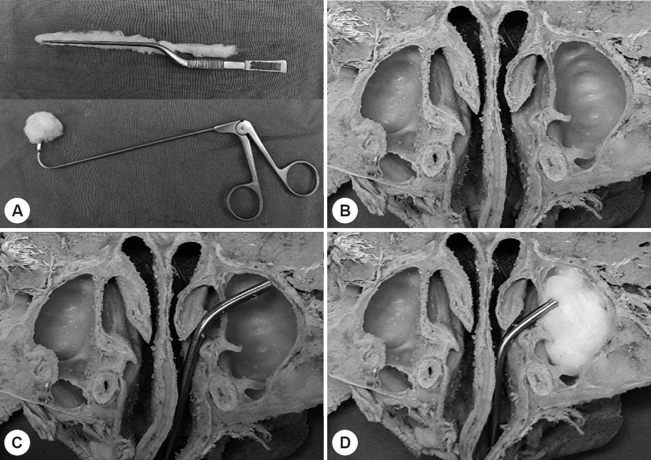
After made a middle meatus antrostomy (MMA), simulation was continued with axially cut cadaver heads to confirm the exact surgical procedure and thoroughly evaluate mucosal injury for paired comparison of cotton pledget and gauze. It was initiated after placing the bulk of adjuvant material in the middle meatus. The adjuvant material was then pushed antero-latero-inferiorly using ball-tip curved suction or 90-degree giraffe forceps (Fig. 1). The procedure was continued until the bulk fully touched the alveolar recess of maxillary sinus. If the surgeon pushes the bulk of the gauze laterally and inferiorly as proven in previous reports, the fungal material will move out toward the maxillary antrostomy site [10,11]. The same procedure was performed with two techniques in random sequence: antibiotic-vaseline soaked cotton pledget and saline soaked gauze.
Easiness in surgical removal and degree of mucosal injury were evaluated by a junior surgeon who was not an author of this article. The easiness of placing adjuvant material into the alveolar recess of maxillary sinus was evaluated using a 10-cm visual analogue scale (VAS). In addition, longest diameters of mucosal tears were summed to evaluate mucosal trauma. The purpose of such evaluation was to find easier and less traumatic adjuvant material for endoscopic fungal ball removal in patients.
Patients
Clinical analysis was focused on the evaluation of efficacy of using cotton pledget technique. Retrospective chart reviews of 88 patients with fungal sinusitis were performed. All 88 patients underwent endoscopic sinus surgery from August 2010 to July 2016 in a tertiary hospital. Fifty-two patients who underwent fungal ball removal using cotton pledget technique were assigned into ‘cotton pledget group’. On the other hand, 36 patients who underwent fungal ball removal without adjuvant material were assigned into the control group. Demographic factors, underlying diseases, preoperative Lund-Mackay (LM) scores, follow-up durations, type of surgeries, types of anesthesia, and incomplete removal rates of both groups were analyzed. All procedures performed in studies involving human participants were in accordance with the ethical standards of the institutional and/or national research committee and with the 1964 Helsinki declaration and its later amendments or comparable ethical standards (IRB number: CNUH-2017-198).
Surgical methods for conventional fungal ball removal
Surgery was performed under local anesthesia or general anesthesia. Gauze strip soaked with a mixture of 4% lidocaine and 1:100000 epinephrine for reducing mucosal swelling was inserted through middle meatus. Sphenopalatine ganglion and anterior ethmoid block were performed with 1% lidocaine with 1:100000 epinephrine with 23G needle.
To provide transnasal corridor to fungal material, an endoscopic middle meatal antrostomy (MMA) was made following the conventional endoscopic sinus surgery technique [12]. An uncinectomy was performed using a sickle knife or a cottle elevator. The natural ostium of maxillary sinus was enlarged and fungal material was identified using 30º and 70º endoscopy. After adequate antrostomy was made, fungal material was removed via MMA using ball-tip curved suction or 90-degree giraffe forceps in both groups. Patients with antral puncture were excluded from this study.
Surgical methods for cotton pledget group
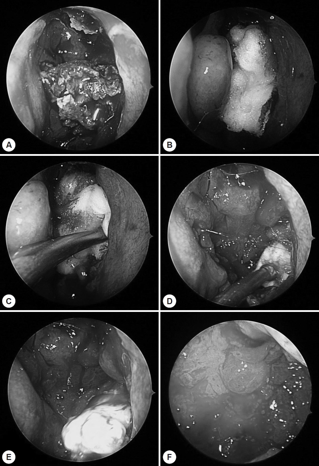
Vaseline coated cotton pledgets were packed at the antrostomy site (Fig. 2, 3). Using ball-tip curved suction, packed adjuvant material into the nasal cavity. In the nasal cavity, the packed adjuvant material usually formed a ball-shaped bulk. As the ball-shaped cotton bulk occupying the maxillary sinus, cheese-like fungal material spilled out from the maxillary sinus to the middle meatus via the antrostomy site. If it was not enough, the operator pushed the adjuvant material anterolaterally along the lateral wall of maxillary sinus using 90º frontal sinus recess giraffe forceps (Fig. 1). The remnant fungal material was then pushed out to the middle meatus.
Statistical analysis
All values are expressed as numbers or means±SD. In cadaveric study, significant differences between the gauze group and the cotton pledget group were determined with Wilcoxon signed ranked test. In clinical study, significant differences between the control group and the cotton pledget group were determined with Fisher’s exact test or Student’s t-test. Statistical significance was set at p<0.05.
RESULTS
Cadaveric study
Mean VAS scores for easiness of gauze and cotton pledget as adjuvant materials were 3.20±1.03 and 4.90±1.37, respectively, with cotton pledget showing significantly (p= 0.011) higher easiness (Table 1). Sizes of mucosal tears in gauze and cotton pledget groups were 3.50±5.06 and 0.50± 1.58, respectively. However, the difference of mucosa tears size between the two groups was not statistically significant (p=0.068).
Clinical study
Mean age of the study population was 65.64±10.80 years. Gender, age, prevalence of hypertension and diabetes, site of disease, or preoperative Lund-Mackay score was not significantly different between the control group and the adjuvant group (Table 2). There were no significant differences in follow up duration or types of surgery between the groups either. However, the adjuvant group showed significantly (p=0.010) lower incomplete removal rate or remnant fungal material than the control group (Table 1).
DISCUSSION
Fungal ball is non-invasive mass-like material commonly found in the elderly [13]. It is more prevalent in hosts with normal immune response. Signs and symptoms of fungal balls are often non-specific and indolent in early stage [14]. The most common symptoms are purulent nasal discharges and nasal obstructions often accompanied by polyp or sinusitis [15,16]. Lesions of cheese-like material or mucopurulent discharge in maxillary sinus during surgery may also occur [17].
Diagnosis of fungal ball can be done by computed tomography (CT) based on characteristics such as increased shading, micro-calcifications, heterogeneous opacifications, and metal-dense spot of the sinus [7,17]. Magnetic resonance images (MRI) can also be used for its diagnosis. T2 MRI images of the fungal mass show marked hypo-intense signal due to iron, magnesium, manganese, and calcium. Therefore, fungal ball has more characteristic features on MRI than that on CT [18].
Treatment of choice for fungal sinusitis is surgical removal without using antifungal agents. However, there is controversy regarding surgery techniques. Before the advent of endoscopic surgical technique for sinus disease (ESS), Caldwell-Luc operation was the mainstay for fungal removal. However, after the advent of ESS technique, endoscopic approaches for fungal sinusitis has gained more popularity than Caldwell-Luc operation [19]. Costa el al. have suggested that endoscopic MMA is the gold standard for fungal ball while a Caldwell-Luc approach has to be reserved for selected cases such as fungal ball of alveolar recess of maxillary sinus [19]. Although endoscopic approach is a good method, it is hard to remove fungal materials of alveolar recess of maxillary sinus by endoscopic approach. Additional antral punctures are performed frequently. However, antral puncture is not a perfect solution in terms of its morbidity [4,5]. It could cause facial swelling, gingival numbness, decreased facial sensation, and wound infection [4].
In this study, we proposed antibiotic-vaseline soaked cotton pledget as an adjuvant material for fungal ball removal instead of antral puncture. Saline soaked gauzes could also be used as adjuvant material [10]. Saline soaked gauze technique was a great idea. We also had experience in using gauze as an adjuvant material for endoscopic maxillary sinus wall reduction [8]. It is a great material for force delivery and stability. However, we thought that the movement of vaseline-coated cotton could be smoother and softer than gauze in the maxillary sinus. Therefore, comparative cadaveric study was performed to select proper adjuvant material. Cadaveric study showed the superiority of vaseline-coated cotton pledget in term of easiness (Table 1). In addition, the degree of mucosal damage caused by cotton pledget was less than that caused by gauze. Although, the different between the two was not statistically significant (Table 1), we should take into account that a small number of dissected cadavers were used in the present study
We used antibiotic-vaseline coated cotton pledget instead of performing antral puncture in this study. Antibiotic-vaseline coated cotton pledget allowed gentle detachment of cheese-like fungal materials from the alveolar recess of maxillary sinus. Its treatment result of the cotton pledget group was significantly better than that of the conventional gauze group. Four patients in the cotton pledget group had remnant fungal material. Three out of these four cases revealed a small amount of remnant fungal material in the early postoperative period within two weeks. All fungal materials in these cases were already detached from maxillary sinuses and easily removed by using a curved suction tip at our outpatient department (OPD). On the contrary, remnant fungal debris in the control group could not be removed easily. All five cases required reoperations.
Although this study has a limitation due to its retrospective, nonrandomized, and non-placebo-controlled study design, our study results showed that fungal ball removal using antibiotic-vaseline soaked cotton pledget was not only easier and time-saving but also an effective in terms of complete removal compared to that using the conventional fungal ball removal procedure.

 PDF Links
 PubReader
 ePub Link
 Full text via DOI
 Download Citation
 Print
Print


